Aleksey Kudreyko1,2,3,* and Vladimir Chigrinov3
1 Department of General Physics, Ufa University of Science and Technology, Ufa 450076, Russia
2 Department of Medical Physics and Informatics, Bashkir State Medical University, Ufa 450008, Russia
3 Microplastics Research Center, Yaroslav-the-Wise Novgorod State University, Velikiy Novgorod 173003, Russia; eechigr2023@gmail.com
* Correspondence: akudreyko@uust.ru
The pervasive contamination of aquatic environments by microplastic particles necessitates the development of rapid, cost-effective and field-deployable detection methodologies to complement established but laboratory-bound spectroscopic techniques such as Fouriertransform infrared and Raman microscopy. The demand for field-suitable methods with a broad accessibility comes from researchers themselves. In this review we systematically examine recent advances in optical methods for microplastics identification with a particular emphasis on birefringence as a key diagnostic feature of partially crystalline synthetic polymers. In particular, we analyze three complementary technological directions: liquid crystal-based sensors that exploit orientational order disruptions at interfaces for label-free microplastics detection; polarization holographic imaging combined with machine learning for high-throughput particle classification; and on-chip polarization light microscopy enabling compact and portable analyzing systems. Liquid crystal platforms demonstrate exceptional sensitivity to submicron particles and enable real-time visualization of microplastics aggregation at aqueous interfaces, though they currently lack polymer-specific chemical identification. Conversely, smart polarization holography integrated with Stokes polarimetry and deep learning algorithms achieves over 90% accuracy in distinguishing microplastics from natural particles while processing up to 10,000 particles per minute. Emerging on-chip polarized light microscopy offers a pathway toward miniaturized, lowcost devices suitable for field applications. By synthesizing insights from foundational studies, this review identifies convergent interdisciplinary trends—particularly the integration of artificial intelligence with multimodal optical imaging—and outlines persistent challenges including standardization, interference from natural organic matter, and the transition from laboratory prototypes to robust field-deployable instruments. The systematization of birefringence-based approaches aims to guide future research towards integrated monitoring systems capable of addressing water quality concerns.
Keywords: microplastics; liquid crystal sensor; environmental monitoring; polarization imaging; polarization light microscopy; in situ detection; machine learning
1. Introduction
In situ detection and identification of microplastic (MP) particles from water represent a global problem. These synthetic polymer particles, ranging in size from 1 μm to 5 mm in length and originating from the fragmentation of macroplastics wastes or intentional manufacturing, exhibit exceptional persistence and ubiquitous prevalence across all environmental compartments, from deep-sea sediments to the Tibetian plateau [1,2]. Their small size and high surface area facilitate the adsorption of co-contaminants, while their potential for trophic transfer and the associated risks of inflammatory responses and cellular toxicity underscore their role as vectors of complex ecotoxicological impact [3,4].
Accurate identification and quantitative analysis of MP particles constitute a fundamental stage in risk assessment and the development of mitigation strategies [5]. Traditional protocols based on sedimentation, filtration and subsequent visual identification under a microscope demonstrate low reproducibility and high probability of misidentification, especially within heterogeneous environmental matrices [6]. The application of spectroscopic methods with chemical specificity, e.g., Fourier-transform infrared (FTIR) and Raman spectroscopy have enabled a transition from mere particle counting to polymer identification [7–9]. However, FTIR and Raman spectroscopy face inherent limitations: FTIR microscopy is constrained by diffraction limits (~10–20 μm) and water interference, while Raman analysis can be hampered by fluorescence and requires prolonged acquisition times for statistically representative sampling [8,10]. Furthermore, FTIR and Raman spectroscopy often require complex sample preparation, which limits their application in field conditions during large-scale screening.
The use of lasers as the excitation source in Raman spectroscopy with an optical microscope enhanced the chemical information to a microscopic level (~500 μm). However this method comes with a distinct set of weaknesses, which are not suitable for application in field condition: additives and pigments cause strong fluorescence, masking polymer spectra [11], and it requires a long analysis time for a small sample area.
These analytical challenges have catalyzed the development of next-generation diagnostic platforms seeking to balance the specificity, sensitivity and potential application in field conditions. Fluorescence-based techniques have emerged as powerful tools [12,13]. More sophisticated approaches, such as Fluorescence Lifetime Imaging Microscopy coupled with phasor analysis, create unique lifetime “fingerprints” for the identification and visualization of MPs in complex matrices [14]. At the same time, advanced label-free optical methods are being developed. For instance, smart polarization and spectroscopic holography integrate digital holography with instantaneous Stokes polarimetry, capturing a rich multidimensional dataset (phase, amplitude, polarization) that serves as a proxy for material properties, enabling classification via machine learning [15]. Fluorescent labeling techniques also make the detection of micro/nanoplastics possible, e.g., luminescent metalphenolic networks composed of zirconium ions, tannic acid, and the fluorescent marker rhodamine B. Such labels make plastic particles as small as 50 nm glow under the excitation light [16]. Then, a mobile application analyzes the obtained image and counts the number of fluorescent pixels.
Thus, polymer materials with a partially crystalline structure are of a special interest for this work, because their internal structure and surface properties can be used for the subsequent optical investigation. Furthermore, the integration of the filtration process with neural network-assisted optical detection methods creates hybrid systems for ensuring continuous water quality.
A particularly intriguing class of methods is based on the application of liquid crystals (LCs) as highly sensitive sensor interfaces. Unlike isotropic fluids, nematic LCs possess a long-range orientational order that is highly susceptible to disruption by interfacial events [17]. Colloidal MP particles, upon adsorption at the LC-aqueous interface, induce elastic deformations within an aligned LC layer. This adsorption leads to the formation of characteristic two-dimensional aggregation patterns, which are unique to different polymer types (e.g., polystyrene and polymethyl methacrylate) and can be altered by environmental factors such as ultraviolet weathering. Application of convolutional neural networks (e.g., DeepPolyNet) for image analysis from the patterns enables accurate predictions of Mps composition, concentration and aging state [9].
Our literature review shows that the main efforts of scientists are aimed at increasing the sensitivity of methods (especially for nanoparticles), and the integration of artificial intelligence for cheap automation of monitoring in the field [18]. An investigation of recently published studies in the detection of MP particles shows the presence of interdisciplinary trends and machine learning methods [16,19,20].
The goal of the present review is to systematize recent advances in optical microplastic identification methods with an emphasis on birefringence as a diagnostic feature. Particular attention is paid to the analysis of birefringent structures—both microplastic particles themselves and filter materials with anisotropic morphology—and their role in the development of integrated monitoring and treatment systems. The paper examines the fundamental principles of polymer optical anisotropy and modern polarization imaging methods, including digital holography and hyperspectral polarimetry, and the potential for applying machine learning methods to automatic particle classification based on multimodal optical features. By synthesizing the insights from recent foundational studies, this work highlights convergent interdisciplinary trends with an emphasis on crystalline structure as a diagnostic feature of microplastic particles. Despite these promising interdisciplinary trends, the current study does not discuss how organic or inorganic crystalline matrices may influence the results (risk of false positives); the influence of matrix effects (e.g., detergents, formation of microplastic aggregates) on detection performance; the response of the method to aging effects and changes in microplastics crystallinity is not addressed; and the limit of detection in terms of particle concentration was not discussed. However, for rapid initial assessment of pollution and monitoring of MPs accumulation, the described optical methods represent an affordable solution.
2. Detection and Visualization Methods of Microplastics
The “gold standard” methods (Section 1) allow simultaneous measurements of particle size, shape, quantity, chemical composition and aging [21]. However, each of these methods has its own constraints. As a result, various modifications of the FTIR and Raman spectroscopy technologies were introduced.
FTIR microscopy combines Fourier Transform Infrared spectroscopy with optical microscopy to provide molecular identification and chemical mapping of samples as small as 5–50 μm due to diffraction constraints [8,10]. This technique scans a filter containing particles and creates “chemical fingerprints” by identifying the polymer at each point by using its IR spectrum. The transition from slow single-point scanning to fast imaging using focal plane array detectors enables the analysis of large filter areas with the spatial resolution (up to ~5–20 μm) within a reasonable time. Recent research advances report that deep convolutional neural networks can extract and recognize complex spectral patterns with 100% of accuracy in classifying aged commercial microplastics samples [9]. However, this method is limited by a spatial resolution of ~10–20 μm. This fact hinders the analysis of small (<10 μm) MPs. This method is also sensitive to moisture, i.e., the sample must be dehydrated.
Raman Spectroscopic Imaging is based on the inelastic scattering of monochromatic light and offers a spatial resolution of less than 1 μm, which is critical for the identification of submicron particles [22]. This method is traditionally used for the analysis of particles in water. The fluorescence can be reduced if the exposing laser radiation has a relatively long wavelength (e.g., 785 nm). A disadvantage of this method is that the aperture of the laser beam ranges within a submicron range. As a result, the measurement time is long, and there is also a risk of sample degradation due to the laser power. There is a lack of standardized protocols and comprehensive spectral libraries that include common additives and aged plastics, making results difficult to compare across studies.
Hybrid methods include surface-enhanced Raman spectroscopy [22], hyperspectral imaging (HSI) [23] and fluorescence-based techniques [12]. Surface-enhanced Raman spectroscopy represents a promising method for the analysis of nanoplastics. Metallic nanostructures dramatically enhance the weak Raman signal, enabling the detection of particles tens of nanometers in size. Optimal nanostructures and sample preparation protocols are under intensive investigation. Hyperspectral imaging is used in the near-infrared range and is ideal for complex environmental samples with mixed or aged plastics [24]. Thus, it is a rapid method for the preliminary screening and sorting of particles, but its chemical information is limited.
Fluorescence-based techniques use specific fluorescent dyes (e.g., the hydrophobic dye Nile Red) that set hydrogen bonding interactions with MPs [25]. Key mechanisms for Mps detection include: hydrophobic absorption, hydrogen and van der Waals interactions and the restriction of intramolecular rotation. Fluorescence-based methods have a number of advantages over FTIR and Raman spectroscopy: the time of analysis ranges within minutes and hours, and they offer high sensitivity and the ability for field applications. Fluorescence methods are successfully used to detect microplastics in various water bodies: wastewater, freshwater and seawater [26,27].
The main challenges that need to be addressed include the elimination of false-positive signals from natural organic matter, the standardizing of protocols, the developing methods for clay soils and the creation of portable systems for continuous monitoring. Further development of detection technologies, combined with advances in chemical synthesis, nanotechnology, and data analysis, will enable the creation of reliable tools for assessing microplastics pollution and minimizing the associated risks. Below, we highlight a number of advanced MPs identification methods based on birefringence and potentially useful for field applications.
3. Liquid Crystal Sensors
Traditional methods of MPs detection are accurate, but require time-consuming sample preparation steps and laboratory conditions, which make operational monitoring in the field impossible. Meanwhile, the patterns of MPs particles self-organization at liquid crystal–aqueous interfaces depend on their chemical nature. This makes it possible to analyze the composition of complex mixtures without resorting to expensive equipment [18].
Liquid crystals consists of highly anisotropic molecules whose orientational order is easily disrupted. A typical sensing process includes the following steps: Sensor preparation. A liquid crystal is deposited on a thin layer of a prepared surface (e.g., a chemically modified gold or glass substrate, which creates an aligned orientation of the LC molecules) [28].
Sample application. An aqueous sample suspected of containing microplastics is applied to the sensor.
Interaction. Microplastic particles with a hydrophobic surface are adsorbed at the interface between the LC and water. This occurs due to the affinity between hydrophobic polymers and the hydrocarbon chains of the LC molecules.
Optical response. The adsorbed particles locally deform the alignment of LC molecules.
Optical microscopy imaging shows bright and dark spots depending on the microscope settings [29]. Aow concentration (e.g., 20 mg/L) generates discrete chain-like aggregates, whereas a high concentration (e.g., 800 mg/L) induces large, multibranched aggregates and extended networks. A higher concentration of microplastic particles leads to the sensor becoming overwhelmed.
Detection. The change is easily visualized or recorded by using a digital camera and image analysis software.
3.1. Liquid Crystal Microfluidic Channel
The unique properties of LCs can be used to “force” microplastics to self-organize into characteristic patterns with the subsequent classification by fractal dimension analysis.
The method of the Abbot group is based on the physical properties of the LC-water interface [30]. This work demonstrated the possibility of identification of polyethylene (PE) and polystyrene (PS) microplastics, which form distinct 2D patterns at LC interfaces.
The experimental setup includes a microfluidic channel to deliver the colloidal fraction of MPs to the aqueous interface of the LC 5CB (4-cyano-4′-pentylbiphenyl) films. This LC layer is deposited on a transmission electron microscopy (TEM) grid, and it must be coated with octyltrichlorosilane. This manipulation is the basis for the formation of a relief and hydrophobicity to achieve a homeotropic alignment of the LC (orange colored surface Figure 1) [31]. Then the excess of LC must be removed.
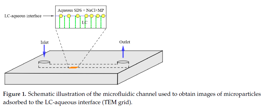
Microplastic particles (e.g., PS) are spontaneously trapped at the water-LC interface. This occurs due to the energetically favorable displacement of the unstable water-LC emulsion by a more stable system, where the particle is located directly at the interface. When a particle enters an LC film, its molecular orientation is distorted. This creates highly visible “flame-like” textures, which can be visualized by using a polarizing microscope. The presence of a low concentration of a surfactant (e.g., 0.025・10−3 M of sodium dodecylsulfate (SDS)) minimizes the aggregation of the microparticles in the bulk aqueous dispersion. The addition of 0.3 M of NaCl initiates the irreversible adsorption of MPs at the LC-aqueous interface.
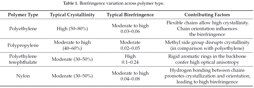
As a result, distinct 2D patterns are assembled. The size of the detected MP particles ranges between 1 and 4 μm. Each independent experiment includes the adjustment of a TEM grid filled with LC and MPs into the channel, and the consequent exposure of the grid (see Figure 1). Then, a number of optical textures can be acquired to show the MPs aggregation within a square of the grid. Polyethylene and PS MPs form distinct 2D patterns at LC interfaces and can be identified by using fractal dimension analysis. The accuracy of classification exceeds 99% and it depends on the concentration of SDS [30]. This difference in features comes from their surface roughness, because PS is an amorphous material with low crystallinity (0–5%) with near-zero birefringence (see also Table 1).
These optical textures are captured on digital camera and analyzed by deep learning models for the concentration of MPs, their size, and their chemical nature (e.g., polymers, polyethylene terephthalate, nylon). The proposed LC-based platform is a multifunctional sensor interface that opens new possibilities for the simple, rapid and sensitive classification of polyethylene and PS with the accuracy of ∼ 99%.
Building upon this platform, the same research group has recently advanced the methodology to address two critical challenges: the analysis of complex mixtures and the influence of environmental weathering [18]. By integrating a convolutional neural network (DeepPolyNet), they demonstrated the ability to identify MPs of polymethyl methacrylate alongside PE and PS, even in the presence of natural organic matter and following UV-induced weathering with the neural network achieving >97% accuracy in composition prediction.
3.2. Liquid Crystal-Infused Porous Polymer Surfaces
The invention of a slippery porous polymer coating impregnated with LC provides a highly sensitive platform for visualization (with the naked eye) and the ability to distinguish amphiphilic substances, i.e., molecules with both water-soluble and fat-soluble parts (e.g., surfactants, lipids, and some proteins) [32]. Thermotropic LCs can be infused into nanoporous polymer membranes to obtain LC-infused surfaces with slippery characteristics in contact with a range of aqueous fluids.
The surface consists of a porous hydrophobic polymer whose pores are filled with LC. This surface is rather slippery for water and many liquids. When a droplet with amphiphilic molecules is in contact with the surface, the molecules interact with the LC 5CB (see Figure 2a). The hydrophobic surface of microplastics (e.g., polyethylene) can directly interact with the LC encapsulated in the pores, causing local changes in its alignment and a different sliding time (Figure 2b). The nature of this effect is that van derWaals interactions play a key role in the binding of the LC and the hydrophobic polymer [33].
Since LCs reflect/refract light differently depending on their orientation, this change is visible with the naked eye as a change in the optical image or color beneath the droplet and at its edges (in the so-called “three-phase contact line”). If we observe this process on slippery liquid-infused porous surfaces (SLIPS), it can be expected that MP particles will not simply aggregate on a flat surface but will interact with the LC impregnation. This interaction can lead to the formation of various optical textures or patterns under a water droplet containing MPs. For example, polyethylene may cause one color/texture change at the point of contact, while PS will exhibit another texture and a different sliding time. The sliding direction is labeled by down arrows in Figure 2b.
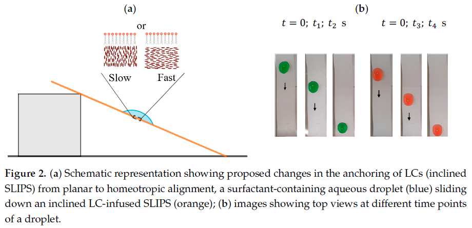
The proposed system requires only a stopwatch and a treated surface (SLIPS) to observe the results. The device reacts to very low concentrations (≥1 nM) of amphiphiles. An array of different SLIPS (with different LC or porous structures) will have different responses to plastic types. The results show that this method is simple and uses inexpensive components, which makes it suitable for work in the field. The practical value of this method lies in the ability to precisely control the overall interaction force at the phase boundary and observe the corresponding changes.
We also add a critical remark on LC sensors. In comparison with the FTIR/Raman methods, LC sensors do not directly measure chemical composition, and their classification is based on morphology and interfacial interactions, which may be ambiguous.
4. High-Throughput Microplastics Assessment
Recent progress in data analysis allows researchers to extract data from MPs assessments [34,35]. For example, the combination of polarization holographic imaging (PHI) and deep-learning methods enables the dynamic analysis of water flow [36–38].
Assuming that the examined polymer materials have a partially crystalline structure, one can apply Stokes parameter analysis for MPs identification. Usually, a Stokes polarimeter is used for this purpose. It is an optical device that captures the four Stokes parameters (see Figure 3a). Each Stokes parameter evaluates specific polarization types over their orthogonal counterparts, i.e., the predominance of linear horizontal (0 degree) over vertical (90 degree) polarization (S1), predominance of linear horizontal (+45 degree) over vertical (−45 degree) polarization (S2), predominance of right-circular (RCP) over left-circular (LCP) polarization (S3) (see Figure 3b) and S0 quantifies the total intensity of the optical beam [39]. Simultaneous measurement of the Stokes parameters enables the analysis of the particles in the flow (dynamic monitoring). In addition, a polarization camera allows researchers to find two basic parameters of the image: (i) the degree of linear polarization (DoLP) and (ii) the angle of linear polarization (AoP), see expressions (1):
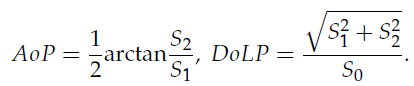
The data from the polarization camera is fed directly into neural networks. This enables us to carry out unique and non-destructive tests on the microstructural evolution of polymers during aging, revealing how molecular order and internal stress change over time. This device offers insights into degradation mechanisms that are not accessible through other optical methods.
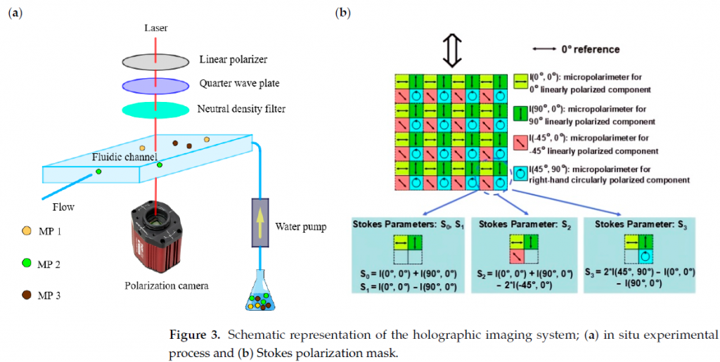
The physical basis of this identification method is as follows: a sample liquid with microplastic particles flows through the device. A laser beam illuminates the particles and creates holographic images. A polarization filter enables information to be extracted from the hologram about how the particle changes the polarization of light. This is a key feature for distinguishing synthetic birefringent polymers from natural particles (such as organic detritus or sand), which are typically non-birefringent. Different MPs materials have a certain optical “fingerprint” in the light polarization data map [40,41]. As a result, MP particles can be classified (e.g., plastic, sand, shells, organic debris) in the water flow. The size and shape of the MPs can also be measured. The main advantages of the Stokes polarimeter are high throughput (i.e., 10 k particles are analyzed per minute), high work efficiency, and an AI-assisted option to distinguish particle types; however, the limit of detection in terms of particle concentration is unknown.
The system rapidly counts microplastic particles in real water samples. It effectively distinguished microplastics from natural particles with over 96% accuracy based on their polarization fingerprints. The high classification accuracy stems from the wide range of birefringence values across polymer types (Table 1), which creates distinct polarization fingerprints detectable by Stokes polarimetry. The method allows particle size determination and primary classification by polymer type (e.g., polyethylene terephthalate, PS, polyethylene).
Thus, polarization holographic imaging is a breakthrough, high-throughput tool for microplastic screening. It combines high speed, AI-assisted automation and the ability to distinguish anthropogenic from natural particles, making it highly promising for large-scale environmental monitoring and research.
5. Polarization-Light Microscopy
Almost all microplastics are formed from synthetic polymers. This fact results in birefringence Δn that ranges within 0.02–0.24 (see Table 1). Note that the birefringence of polyethylene terephthalate with one benzene ring achieves 0.24. This is because the birefringence value is explained not only by the presence of the benzene ring, but also by the high anisotropy of the polarizability of the ester group (–CO–O–), and, most importantly by the perfect trans-conformation of the main chain, which aligns all these polarizable elements strictly parallel to the orientation axis [42].
In the visible spectrum λ = 380 . . . 760 nm, the characteristic length is given by d = λ/Δn. This allows the detection of MP particles with the size of 10–20 μm [43].
The structural specificity of MPs exhibit polarized light behavior, such as reflection, refraction, and scattering, which allows them to be distinguished from other types of materials [44,45]. Such simple phenomena are not just a list of properties, but a guide for the fabrication of inexpensive and easy-to-use systems for the highly sensitive analysis of organic samples (cells, tissues, biomolecules and microplastic) without the need for complex and bulky microscopes [46].
Although monosodium urate crystals and MP particles may seem at first glance to be completely different objects, their comparison is scientifically revealing. It allows us to understand how nature and anthropogenic pollution create structurally similar but biologically opposite problems. The formation of monosodium urate crystals with a highly ordered internal crystal structure is a complex pathological process that occurs inside joints with pH ∼7.4 [47,48]. Unlike simple precipitation, monosodium urate crystallization proceeds through the formation of fibril-like amorphous subunits, which then agglomerateand transform into needle-shaped crystals.
Typical cross-polarized experiments show the possibility of registering a high contrast between birefringent needle-shaped monosodium urate crystals and the non-birefringent background [49]. A distinct feature of monosodium urate crystals is their strong negative birefringence Δn ∼−0.1 [50]. This allows researchers to differentiate monosodium urate crystals from other materials with a crystalline structure.
Figure 4a depicts the polarization imaging setup: a non-polarized illumination source is filtered by a 50 μmpinhole, after which the resulting point source illumination is collected by a lens mounted in a threaded tube. The distance from the lens to the pinhole is adjusted in such a way as to produce a collimated beam. Then the light beam is polarized (LP 1, 120 μm thickness) and travels through a full waveplate (150 μm thickness) positioned at ± 45°, both of which are placed in manual rotating mounts for angle control.
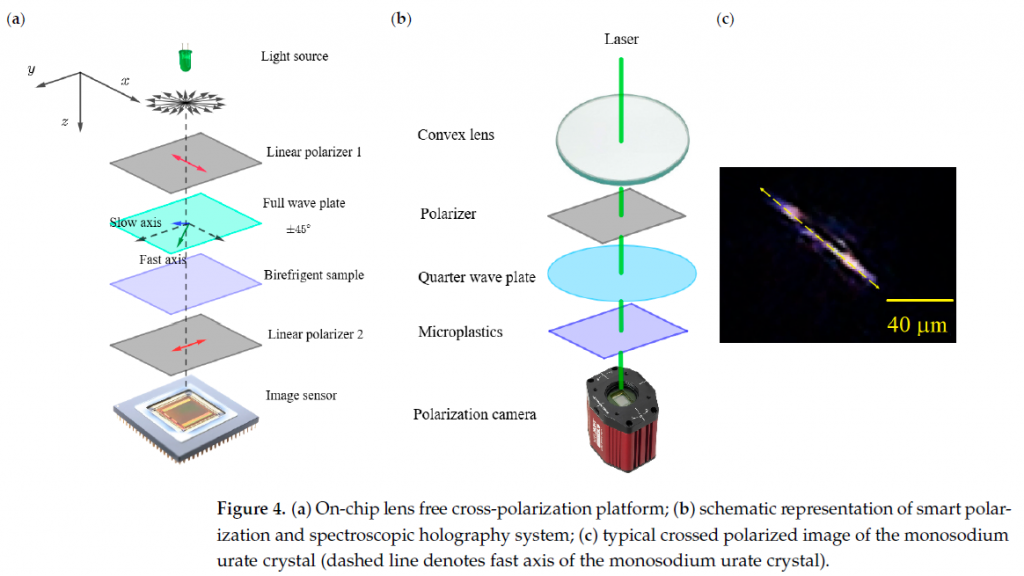
As soon as the LP 1 is introduced, only one polarization interacts with the birefringent sample. The polarized light is split into ordinary and extraordinary beams, which have a phase difference equal to π within the sample’s pathlength. The recombination of light after emerging from the sample results in a phase shift and polarization (since the effective polarization vector is rotated). This effect occurs for all wavelengths. A full wave-plate is used to add or subtract the effective retardance from the sample, depending on its orientation in relation to the polarization of the incident light (± 45°). Then, the linear polarizer 2 reveals a colorful interference pattern that indicates the birefringence of the sample. The image sensor registers this pattern as an image for the subsequent data processing. Rotation of the full waveplate around ± 45° induces different interference color patterns, and colored images can be obtained. The output signal correlates with the quantity of anisotropic structures and its alignment in the sample.
The visualization method was tested on several types of samples: Euglena gracilis, monosodium urate crystals, ascorbic acid crystals and a mica plate. This technique can be further miniaturized and built into a portable, point-of-care/need device for a variety of biological and non-biological samples.
Further transformation of polarization-light microscopy into smart polarization and spectroscopic holography enables to capture multi-dimensional features that include polarization states (S0, S1, S2, S3), phase retardation, etc. [15,46]. Such a combination of these features with machine learning methods does not require the implementation of a spectroscopic system (see Figure 4b). The quarter wave plate changes linear into circular polarization of light. Then, elliptically polarized light interacts with the long polymer chains of the MPs and a colorful interference pattern is observed. A typical image of a monosodium urate crystal without a full waveplate is depicted in Figure 4c.
Here, the polarization camera includes the Stokes polarization mask depicted in Figure 3b [51]. This technique represents a promising tool for addressing pressing environmental issues such as assessing microplastic pollution, identifying its sources, and conducting long-term monitoring of water quality.
6. Discussion
The three birefringence-based approaches examined here—LC sensors, polarization holographic imaging, and on-chip polarization microscopy occupy different but complementary niches in the microplastic detection landscape. LC sensors excel at submicron sensitivity and the real-time visualization of particle aggregation at interfaces. The reliance of LCs on morphological patterns rather than direct chemical signatures makes them most suitable as rapid screening tools prior to spectroscopic confirmation. Conversely, PHI coupled with Stokes polarimetry offers a high throughput (up to 104 particles/min) and polymer-type differentiation via intrinsic birefringence fingerprints, positioning it as a candidate for automated monitoring stations where speed and minimal sample handling are critical. On-chip polarization microscopy provides a low-cost, field-portable architecture, though its current detection limit (10–20 μm) and sensitivity to turbidity require further validation for complex environmental matrices (see Table 2).
A key unresolved question across all three methods is the quantitative impact of UV weathering and biodegradation on birefringence values [52]. As shown in Table 1, crystallinity and polymer chain orientation govern Δn; surface oxidation, chain scission, or secondary crystallization during aging may systematically alter polarization signatures, potentially reducing classification accuracy or causing false negatives. Systematic aging studies with well-characterized reference materials are needed to establish correction factors or retrain machine learning models for environmentally aged microplastics [53].
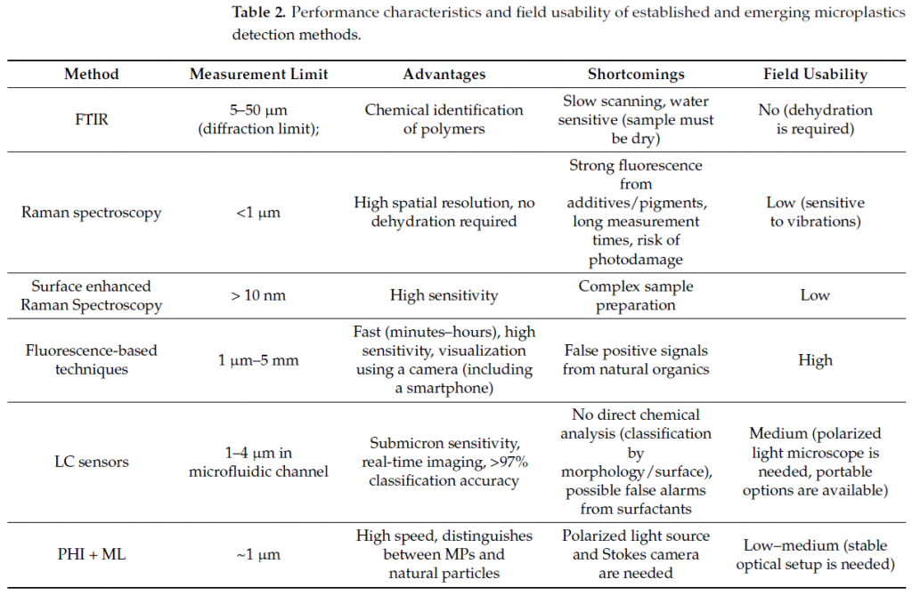
The most promising near-term advance lies in hybrid configurations—for example, using LC-coated microfluidic interface to trap and preconcentrate particles, followed by an on-chip Stokes readout [54]. Such integration would combine the sensitivity of LC alignment disruption with the chemical-specific polarization fingerprints of PHI, potentially overcoming the principal limitation of each method when used alone.
7. Conclusions
Many studies show the need to develop field-appropriate microplastic detection methods. The partially crystalline nature of many synthetic polymers imparts measurable birefringence, which serves as a powerful intrinsic contrast mechanism for optical detection. This review has systematically examined three complementary approaches that exploit birefringence for microplastic identification: LC based sensors, polarization holographic imaging with machine learning and on-chip polarization light microscopy.
LC-based sensors offer an exceptional sensitivity to submicron particles (<1 μm) and enable real-time visualization of microplastic aggregation at aqueous interfaces without fluorescent labeling. When combined with fractal dimension analysis or convolutional neural networks (e.g., DeepPolyNet), they achieve >97% accuracy in distinguishing polymer types even after UV weathering. Their main limitation is the lack of direct chemical specificity—classification relies on interfacial interactions and morphology.
Polarization holographic imaging integrated with Stokes polarimetry and deep learning achieves a high throughput (up to 10 k particles per minute) with >90–96% accuracy in distinguishing microplastics from the natural birefringent background (sand, organic debris). It provides the particle size distribution and polymer-type classification (PET, PS, PE) based on distinct birefringence fingerprints. The technique is label-free and non destructive, but its detection limit in terms of particle concentration and performance in turbid media remain to be systematically evaluated.
On-chip polarization light microscopy represents a pathway toward miniaturized, low-cost devices suitable for field use. Using simple cross-polarized optics without bulky lenses, it can detect birefringent particles down to 10–20 μm and has been successfully demonstrated for monosodium urate crystals—a model birefringent analyte. Adaptation to microplastics requires further validation, but the concept aligns well with the need for point-of-need monitoring.
Despite these advances, several cross-cutting challenges persist. First, the standardization of sample preparation, data acquisition, and reporting is lacking, hindering interlaboratory comparison. Second, natural organic matter, detergents, and microplastic aggregates can produce false-positive signals or alter aggregation patterns. Third, the influence of UV weathering and aging on birefringence values is not yet fully characterized, which may affect long-term reliability. Fourth, LC sensors do not provide chemical identification, and polarization holographic imaging methods require further refinement to reduce false positives in complex environmental matrices.
Looking forward, the integration of these technologies offers promising synergies. Hybrid systems combining LC interface trapping with a Stokes polarimetry readout could enhance both sensitivity and specificity. The incorporation of advanced machine learning (vision transformers, generative models) will likely improve classification robustness with limited training data.
In summary, birefringence-based optical methods provide a powerful, label-free toolkit for microplastic detection that balances speed, cost, and information content. While not yet a replacement for FTIR or Raman spectroscopy in definitive chemical identification, they are uniquely suited for high-throughput screening, initial pollution assessment and continuous environmental surveillance.
Author Contributions: Conceptualization, A.K. and V.C.; writing, editing and overview, A.K. All authors have read and agreed to the published version of the manuscript.
Funding: This study was funded by the Ministry of Science and Higher Education of the Russian Federation (state contract no. 075-15-2025-016, MegaGrant).
Data Availability Statement: The original contributions presented in this study are included in the article. Further inquiries can be directed to the corresponding author.
Conflicts of Interest: The authors declare no conflicts of interest.
References
1. Peng, X.; Chen, M.; Chen, S.; Dasgupta, S.; Xu, H.; Ta, K.; Du, M.; Li, J.; Guo, Z.; Bai, S. Microplastics contaminate the deepest part of the world’s ocean. Geochem. Perspect. Lett. 2018, 9, 1–5. [CrossRef]
2. Zhang, Y.; Gao, T.; Kang, S.; Allen, S.; Luo, X.; Allen, D. Microplastics in glaciers of the Tibetan Plateau: Evidence for the long-range transport of microplastics. Sci. Total Environ. 2021, 758, 143634. [CrossRef]
3. Cao, J.; Xu, R.; Wang, F.; Geng, Y.; Xu, T.; Zhu, M.; Lv, H.; Xu, S.; Guo, M.-Y. Polyethylene microplastics trigger cell apoptosis and inflammation via inducing oxidative stress and activation of the NLRP3 inflammasome in carp gills. Fish Shellfish Immunol. 2023, 132, 108470. [CrossRef]
4. Gautam, R.; Jo, J.; Acharya, M.; Maharjan, A.; Lee, D.; Kc, P.B.; Kim, C.; Kim, K.; Kim, H.; Heo, Y. Evaluation of potential toxicity of polyethylene microplastics on human derived cell lines. Sci. Total Environ. 2022, 838, 156089. [CrossRef] [PubMed]
5. Ibrahim, N.; Rahman, A.M.N.A.A.; Shafiq, M.D.; Lockman, Z.; Jaafar, M.; Kameda, Y. Microplastic pollution: Sources, degradation mechanisms, analytical advances, and mitigation strategies for environmental sustainability. Rev. Environ. Contam. Toxicol. 2025, 263, 27. [CrossRef]
6. Fernandez-Gonzalez, V.; Andrade-Garda, J.M.; Lopez-Mahia, P.; Muniategui-Lorenzo, S. Misidentification of PVC microplastics in marine environmental samples. TrAC Trends Anal. Chem. 2022, 153, 116649. [CrossRef]
7. Ribeiro-Claro, P.; Nolasco, M.M.; Araujo, C. Characterization of microplastics by Raman spectroscopy. Compr. Anal. Chem. 2017, 75, 119–151.
8. Chen, Y.; Wen, D.; Pei, J.; Fei, Y.; Ouyang, D.; Zhang, H.; Luo, Y. Identification and quantification of microplastics using Fourier-transform infrared spectroscopy: Current status and future prospects. Curr. Opin. Environ. Sci. Health 2020, 18, 14–19. [CrossRef]
9. Zeng, G.; Ma, Y.; Du, M.; Chen, T.; Lin, L.; Dai, M.; Luo, H.; Hu, L.; Zhou, Q.; Pan, X. Deep convolutional neural networks for aged microplastics identification by Fourier transform infrared spectra classification. Sci. Total Environ. 2024, 913, 169623 [CrossRef]
10. Veerasingam, S.; Ranjani, M.; Venkatachalapathy, R.; Bagaev, A.; Mukhanov, V.; Litvinyuk, D.; Mugilarasan, M.; Gurumoorthi, K.; Guganathan, L.; Aboobacker, V. Contributions of Fourier transform infrared spectroscopy in microplastic pollution research: A review. Crit. Rev. Environ. Sci. Technol. 2021, 51, 2681–2743. [CrossRef]
11. Azari, A.; Ronsmans, S.; Vanoirbeek, J.A.; Hoet, P.H.; Ghosh, M. Challenges in Raman spectroscopy of (micro) Plastics: The interfering role of colourants. Environ. Pollut. 2024, 363, 125250. [CrossRef]
12. Bhavsar, P.S.; Devre, P.V.; Sawat, A.H.; Patil, A.S.; Nille, O.S.; Naik, V.M.; Dongare, P.R.; Gunjal, D.B.; Panja, S.K.; Gore, A.H. Applications of fluorescence-based techniques for microplastics and nanoplastics analysis. In Fluorescence Spectroscopy in Analytical Chemistry; Elsevier: Amsterdam, The Netherlands, 2026; pp. 531–552.
13. Merck, N.; Otto, J.; Schaeper, M.; Damaschke, N. A non-contact in situ approach for detecting fluorescent microplastic particles in flowing water using fluorescence spectroscopy. Environ. Sci. Adv. 2026, 5, 206–220. [CrossRef]
14. Monteleone, A.; Schary, W.; Wenzel, F.; Langhals, H.; Dietrich, D.R. Label-free identification and differentiation of different microplastics using phasor analysis of fluorescence lifetime imaging microscopy (FLIM)-generated data. Chem.-Biol. Interact. 2021, 342, 109466. [CrossRef] [PubMed]
15. Zhu, Y.; Li, Y.; Huang, J.; Lam, E.Y. Smart polarization and spectroscopic holography for real-time microplastics identification. Commun. Eng. 2024, 3, 32. [CrossRef]
16. Ye, H.; Zheng, X.; Yang, H.; Kowal, M.D.; Seifried, T.M.; Singh, G.P.; Aayush, K.; Gao, G.; Grant, E.; Kitts, D. Cost-effective and wireless portable device for rapid and sensitive quantification of micro/nanoplastics. ACS Sens. 2024, 9, 4662–4670. [CrossRef]
17. Kudreyko, A.; Chigrinov, V. Optical Biosensing of Polarized Light. Crystals 2023, 13, 1680. [CrossRef]
18. Mukherjee, F.; Shi, A.; Latasa, L.A.; You, F.; Abbott, N.L. Liquid crystal–driven interfacial ordering of colloidal microplastics: Advancing microplastic characterization below the macroscale. Sci. Adv. 2025, 11, eady1167. [CrossRef]
19. Guo, M.; Guo, B.; He, S.; Zhou, Z.; Huang, H.; Li, P. Computational polarimetric holography for efficient microplastic classification via a lightweight wavelet-enhanced vision transformer. Opt. Express 2026, 34, 9807–9827. [CrossRef] [PubMed]
20. Nyakuchena, M.; Juntunen, C.; Sung, Y. Deep-learning-assisted near-infrared hyperspectral imaging for microplastic classification. Powder Technol. 2025, 457, 120933. [CrossRef]
21. Zhu, Y.; Li, Y.; Huang, J.; Zhang, Y.; Ho, Y.W.; Fang, J.K.H.; Lam, E.Y. Advanced optical imaging technologies for microplastics identification: Progress and challenges. Adv. Photonics Res. 2024, 5, 2400038. [CrossRef]
22. Levermore, J.M.; Smith, T.E.; Kelly, F.J.;Wright, S.L. Detection of microplastics in ambient particulate matter using Raman spectral imaging and chemometric analysis. Anal. Chem. 2020, 92, 8732–8740. [CrossRef]
23. Faltynkova, A.; Johnsen, G.;Wagner, M. Hyperspectral imaging as an emerging tool to analyze microplastics: A systematic review and recommendations for future development. Microplast. Nanoplast. 2021, 1, 13. [CrossRef]
24. Faltynkova, A.; Wagner, M. Developing and testing a workflow to identify microplastics using near infrared hyperspectral imaging. Chemosphere 2023, 336, 139186. [CrossRef] [PubMed]
25. Sancataldo, G.; Avellone, G.; Vetri, V. Nile Red lifetime reveals microplastic identity. Environ. Sci. Process. Impacts 2020, 22, 2266–2275. [CrossRef] [PubMed]
26. Nicolai, E.; Pizzoferrato, R.; Li, Y.; Frattegiani, S.; Nucara, A.; Costa, G. A new optical method for quantitative detection of microplastics in water based on real-time fluorescence analysis. Water 2022, 14, 3235. [CrossRef]
27. Meng, X.; Chen, S.; Li, D.; Song, Y.; Sun, L. Identification of marine microplastics based on laser-induced fluorescence and principal component analysis. J. Hazard. Mater. 2024, 465, 133352. [CrossRef] [PubMed]
28. Oladepo, S.A. Development and application of liquid crystals as stimuli-responsive sensors. Molecules 2022, 27, 1453. [CrossRef] [PubMed]
29. Carlton, R.J.; Hunter, J.T.; Miller, D.S.; Abbasi, R.; Mushenheim, P.C.; Tan, L.N.; Abbott, N.L. Chemical and biological sensing using liquid crystals. Liq. Cryst. Rev. 2013, 1, 29–51. [CrossRef]
30. Mukherjee, F.; Shi, A.; Wang, X.; You, F.; Abbott, N.L. Liquid crystals as multifunctional interfaces for trapping and characterizing colloidal microplastics. Small 2023, 19, 2207802. [CrossRef]
31. Han, T.; Kim, J.H.; Kim, Y.D.; Ahn, D.J.; Lim, D.-K. Solution-based one-step preparation of three-dimensional self-assembled octadecyl silica nanosquare plate and microlamella structures for superhydrophobic and icephobic surfaces. Langmuir 2021, 37, 5886–5894. [CrossRef]
32. Agarwal, H.; Nyffeler, K.E.; Manna, U.; Blackwell, H.E.; Lynn, D.M. Liquid Crystal-Infused Porous Polymer Surfaces: A “Slippery” Soft Material Platform for the Naked-Eye Detection and Discrimination of Amphiphilic Species. ACS Appl. Mater. Interfaces 2021, 13, 33652–33663. [CrossRef] [PubMed]
33. Wang, H.; Evans, D.; Voelcker, N.H.; Griesser, H.J.; Meagher, L. Modulation of substrate van der Waals forces using varying thicknesses of polymer overlayers. J. Colloid. Interface Sci. 2020, 580, 690–699. [CrossRef] [PubMed]
34. Visentin, E.; Niero, G.; Benetti, F.; O’Donnell, C.; De Marchi, M. Assessing microplastic contamination in milk and dairy products. npj Sci. Food 2025, 9, 135. [CrossRef]
35. Lee, S.; Jeong, H.; Hong, S.M.; Yun, D.; Lee, J.; Kim, E.; Cho, K.H. Automatic classification of microplastics and natural organic matter mixtures using a deep learning model. Water Res. 2023, 246, 120710. [CrossRef]
36. Li, Y.; Zhu, Y.; Huang, J.; Ho, Y.-W.; Fang, J.K.-H.; Lam, E.Y. High-throughput microplastic assessment using polarization holographic imaging. Sci. Rep. 2024, 14, 2355. [CrossRef]
37. Zou, S.; Ju, H.; Zhang, J.Water quality management in the age of AI: Applications, challenges, and prospects. Water 2025, 17, 1641. [CrossRef]
38. Lu, R.; Lin, Z.; Dong, J.; Nan, H.; Jin, Q.; Wu, X. Automated multi-parameter characterization of microplastics using polarization digital holographic microscopy. Appl. Opt. 2026, 65, 3473–3482. [CrossRef]
39. Berry, H.G.; Gabrielse, G.; Livingston, A. Measurement of the Stokes parameters of light. Appl. Opt. 1977, 16, 3200–3205. [CrossRef]
40. Bianco, V.; Pirone, D.; Memmolo, P.; Merola, F.; Ferraro, P. Identification of microplastics based on the fractal properties of their holographic fingerprint. ACS Photonics 2021, 8, 2148–2157. [CrossRef]
41. Valentino, M.; Behàl, J.; Pirone, D.; Memmolo, P.; Miccio, L.; Bianco, V.; Ferraro, P. On the use of machine learning for microplastic identification from holographic phase-contrast signatures. In Proceedings of the Multimodal Sensing and Artificial Intelligence: Technologies and Applications III; SPIE: Bellingham,WA, USA, 2023; pp. 216–223.
42. Kunugi, T.; Shiratori, K.; Uematsu, K.; Hashimoto, M. Estimation of intrinsic birefringence of poly (ethylene terephthalate) crystal. Polymer 1979, 20, 171–174. [CrossRef]
43. Kalaronis, D.; Ainali, N.M.; Evgenidou, E.; Kyzas, G.Z.; Yang, X.; Bikiaris, D.N.; Lambropoulou, D.A. Microscopic techniques as means for the determination of microplastics and nanoplastics in the aquatic environment: A concise review. Green Anal. Chem. 2022, 3, 100036. [CrossRef]
44. Pierro, M.P.; Valentino, M.; Ducoli, S.; Miccio, L.; Bianco, V.; Federici, S.; Ferraro, P. Discerning Pristine and Aged True-to-life Microplastics through Polarization-resolved Digital Holography and Machine Learning. J. Environ. Chem. Eng. 2026, 14, 122875. [CrossRef]
45. Genc, S.; Icoz, K.; Erdem, T. Numerical analysis and experimental verification of optical scattering from microplastics. R. Soc. Open Sci. 2023, 10, 230586. [CrossRef]
46. Pedrosa, T.d.L.; de Araujo, R.E.; Wachsmann-Hogiu, S. On-Chip Polarization Light Microscopy. Biosensors 2025, 15, 79. [CrossRef]
47. Rodriguez-Navarro, C.; Elert, K.; Ibanez-Velasco, A.; Monasterio-Guillot, L.; Andres, M.; Sivera, F.; Pascual, E.; Ruiz-Agudo, E. Unraveling the pathological biomineralization of monosodium urate crystals in gout patients. Commun. Biol. 2024, 7, 828. [CrossRef]
48. Martillo, M.A.; Nazzal, L.; Crittenden, D.B. The crystallization of monosodium urate. Curr. Rheumatol. Rep. 2014, 16, 400. [CrossRef]
49. Park, S.; Lee, L.E.; Kim, H.; Kim, J.E.; Lee, S.J.; Yoon, S.; Shin, S.; Kang, H.; Park, Y.; Song, J.J. Detection of intracellular monosodium urate crystals in gout synovial fluid using optical diffraction tomography. Sci. Rep. 2021, 11, 10019. [CrossRef]
50. Zhang, Y.; Lee, S.Y.C.; Zhang, Y.; Furst, D.; Fitzgerald, J.; Ozcan, A. Wide-field imaging of birefringent synovial fluid crystals using lens-free polarized microscopy for gout diagnosis. Sci. Rep. 2016, 6, 28793. [CrossRef] [PubMed]
51. Wang, Y.; Su, Y.; Sun, X.; Hao, X.; Liu, Y.; Zhao, X.; Li, H.; Zhang, X.; Xu, J.; Tian, J. Principle and implementation of stokes vector polarization imaging technology. Appl. Sci. 2022, 12, 6613. [CrossRef]
52. Gao, L.; Su, Y.; Mehmood, T.; Wang, Z.; Peng, L.; Zhang, N. UVA-induced weathering of microplastics in seawater: Surface property transformations and kinetics. Front. Mar. Sci. 2025, 12, 1519668. [CrossRef]
53. Li, L.; Xue, B.; Lin, H.; Lan, W.; Wang, X.; Wei, J.; Li, M.; Li, M.; Duan, Y.; Lv, J. The adsorption and release mechanism of different aged microplastics toward Hg (II) via batch experiment and the deep learning method. Chemosphere 2024, 350, 141067. [CrossRef]
54. Valentino, M.; B˘ehal, J.; Bianco, V.; Itri, S.; Mossotti, R.; Dalla Fontana, G.; Battistini, T.; Stella, E.; Miccio, L.; Ferraro, P. Intelligent polarization-sensitive holographic flow-cytometer: Towards specificity in classifying natural and microplastic fibers. Sci. Total Environ. 2022, 815, 152708. [CrossRef] [PubMed]
Disclaimer/Publisher’s Note: The statements, opinions and data contained in all publications are solely those of the individual author(s) and contributor(s) and not of MDPI and/or the editor(s). MDPI and/or the editor(s) disclaim responsibility for any injury to people or property resulting from any ideas, methods, instructions or products referred to in the content.




![Микропластик в воде и пище: [не]осведомленность](/upload/iblock/3a0/h99p373wv55ur266lw4qbbegvp6f8033.jpg)